
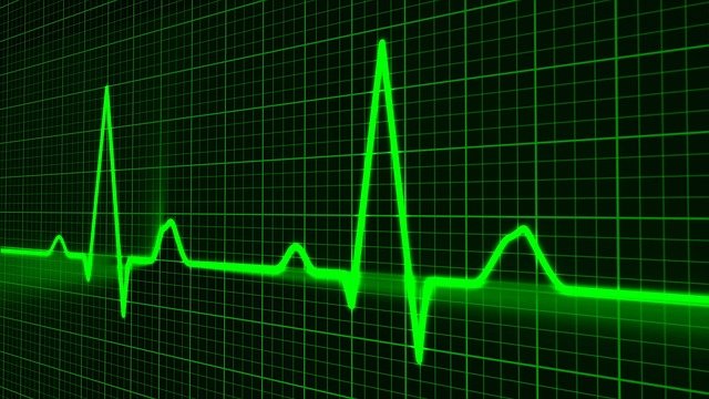
Remember those movie scenes, where they rush in a patient through the emergency room doors of the hospital, and the patient’s heart suddenly stops beating? They give him an electric shock, but there is still a flat line on the computer. They wait for a second, give him another shock, suddenly the monitor shows a wave, and the heart starts beating again. Everyone breathes a sigh of relief. Do you ever wonder, how an electric shock could bring a person back to life? Aren’t we told that electricity is dangerous and should not go poking our fingers into sockets?
Feeling your Heart beat
In 1886, Walter Gaskell showed that our heart is electrical in nature. It is the electrical nature of the heart that allows it to keep pumping blood all day and night. We do not have to tell our heart to beat, hence it is said to be under ‘involuntary control’.
The heart pumps oxygenated blood through blood vessels called arteries to various parts of the body, and deoxygenated blood is collected from the various parts back to the heart through blood vessels called veins. The beating of the heart allows the regular flow or pumping of the blood.
Doctors use a stethoscope to hear the heartbeat. You can feel the pumping of the blood through the heart, by checking your pulse. Most often, the pulse is felt at the carotid artery near the neck or by placing a finger on your wrist near the thumb at the radial artery. One does not use veins to check their pulse, because veins have relatively thin-walled and do not show the pulse. Your heart beats at an average of 72 times per minute and during that time pumps out almost 5 litres of blood!
The Chambers of the heart
Let us take a closer look at how the pumping of the blood happens. The heart has 4 chambers or compartments that are divided into two sides. The left side of the heart has the left atrium and the left ventricle and the right side of the heart has the right atrium and right ventricle. The chambers on the right side, have deoxygenated blood, and the chambers on the left have oxygenated blood. The blood never mixes.

The heart collects deoxygenated blood from the various parts of the body into the right atrium. From there, it sends it to the right ventricle through a valve.
Consider the valve to be like a door that opens only from the inside. Once the blood has entered the ventricle, it will not go back to the atrium due to the valve. From the right ventricle, the blood is pumped out through the pulmonary artery to the lungs to get oxygenated and the oxygenated blood returns back to the heart through the pulmonary vein at the left atrium.
The pulmonary artery is the only artery that carries deoxygenated blood in the body, while the pulmonary vein is the only vein that carries oxygenated blood. This is because, all blood vessels that leave the heart are called arteries, and all blood vessels that bring blood to the heart are called veins. From the left atrium, the oxygenated blood enters the left ventricle, through another valve. Finally, the oxygenated blood leaves the heart through the aorta and supplies the body.
The Electrical Nature of the Heart
The heart is made up of muscle, and the electrical impulse is carried out throughout the heart through it. The electrical impulse originates in a region called the sinoatrial node or the SA node, which is present in the muscular tissue of the right atrium.
The impulse is regularly generated in the SA node and it travels down conduction pathways. The atria of the heart first contract, which pumps the blood into the ventricles. The electrical impulse reaches a region called the AV node or atrioventricular node.
Here, the impulse slows down for a bit, which allows the blood from the atrium to enter the ventricle before the ventricle pumps it out. After the AV node, the electrical impulse is carried forward through a conduction pathway, called the bundle of His, which is between the two ventricles, and finally, the bundle of His divides into the left and right side of the ventricles.
This causes the ventricles to contract and pushes the blood out of the heart. After this, both the atria and the ventricles relax, which allows blood to enter the atria.
External regulation of the Heart
While watching a horror movie in the theatre, or your favorite actor on screen, you feel your heart beating faster. When you exercise, your heart starts beating faster. Apart from the control that the heart has on its own, the nervous system also controls the heart rate.
In conditions, where you are scared, the parasympathetic nervous system directs your heart to beat faster. This allows more blood flow to the extremities of your body like your hands and legs, so that you can react or get away quickly from a scenario.
You may have seen an adrenaline injection given to restart the heart. Adrenaline is the same hormone produced naturally in your body that helps in signaling through your parasympathetic nervous system. On other occasions, when you are at rest or when your sympathetic nervous system is active, your nervous system signals your heart to slow down a bit.
The machine used to give electric shocks to the heart is called a defibrillator. It produces a high energy electric shock which would hopefully return the heart to its normal state in case of cardiac arrest.

It is important to remember that our bodies conduct electricity. So unless you are protected by an insulating material, if you get a high voltage electric shock on your skin, you are likely to burn your skin. In the worst-case scenario, you can die of electrocution.
Storing a Heart for a transplant
Immediately after death has been declared, all the cells in the body use up their remaining energy and within 5 to 6 minutes die entirely. When kept in the cold, the cells of the heart stay alive for about 4 to 6 hours after they have been removed out of a donor’s body when kept in the cold.
The heart may not beat, but the cells of the heart do not die immediately. Just like other organs that are transplanted, they are kept in the cold. In the cold, the cells decrease their metabolic rate, reduce their energy requirements, and hence can stay alive till the run out entirely of energy. Of course, for organ transplants, there is a time period, beyond which the organs are considered too damaged to be transplanted.
Newer techniques, now keep the heart pumping put of the donor’s body with a continuous supply of blood and nutrients.
Your heart muscle works continuously without a break, till the day it stops and you die. It has been seen that the more heart beats, the shorter life span, the animal has. The life span of a rat that has a heart rate of 400 bpm (beats per minute) is around 5 years.
On the other hand the life span of a giant Galapagos turtle of around 8 bpm has a life expectancy of around 177 years.
It has been found that humans who have a higher resting heart rate have a shorter life expectancy than those who have a higher resting heart rate.
Further reading:
Silverman, M. E., Grove, D., & Upshaw Jr, C. B. (2006). Why does the heart beat? The discovery of the electrical system of the heart. Circulation, 113(23), 2775-2781.
https://www.livescience.com/how-long-can-donated-organs-last-before-transplant.html
Read more Articles from Life Sciences
1. Read about the Start Your Own Fruit Fly Culture at Home
2. Read about the Did You Just Spot a Cheetah a Leopard and a Jaguar
3. Read about the The Secret Lives of Tardigrades
4. Read about the Chlorophyll and Haemoglobin – An Unlikely Connection
5. Read about the What is the True Colour of the Coronavirus?
6. Read about the 7 Fascinating Facts About Fruit Flies
About Author
Saunri Dhodi Lobo is pursuing M.Sc in Life Sciences with specialization in Neurobiology. Her interests include writing poetry, going for nature walks and swimming. Currently she is involved in research on Alzheimer’s Disease in fruit flies.
Read all Articles by Saunri Dhodi Lobo
Photos, Vector Graphics & Illustrations Credits
Human heart by By Wapcaplet, Yaddah – Own work, CC BY-SA 3.0, https://commons.wikimedia.org/w/index.php?curid=830246
Defibrillator – The photo was taken in Biomedical Systems Department of MIET, CC BY-SA 3.0, https://commons.wikimedia.org/w/index.php?curid=2122699